Intan Risna/ Tropmed 7/ Case 4
(versi males mentranslasi ^^, I wish I could someday)
INTESTINAL AND UROGENITAL PROTOZOA
Intestinal and luminal protozoa significant to human health include
- Entamoeba histolytica (Amebae)
- Balantidium coli (Ciliates)
- Giardia lamblia and Trichomonas vaginalis (Flagellates)
- Cryptosporidium parvum and Isospora belli (Sporozoa)
AMEBIASIS (amebic dysentery, amebic hepatitis)
Etiology
E. histolytica is the major cause of amebic dysentery.
Epidemiology
0.5 to 50% of the population world wide harbors E. histolytica parasites with the higher rates of infection being in underdeveloped countries. 1 to 3% of the population of the USA are infected. Infection is associated with poor hygiene. Humans are the principal host, although dogs, cats and rodents may be infected.
MorphologyLife cycle
Trophozoite: This form has an ameboid appearance and is usually 15-30 micrometers in diameter, although more invasive strains tend to be larger. The organism has a single nucleus with a distinctive small central karyosome (Figure 1A,B). The fine granular endoplasm may contain ingested erythrocytes (Figure 1C). The nuclear chromatin is evenly distributed along the periphery of the nucleus.Cyst: Entameba histolytica cysts are spherical, with a refractile wall; the cytoplasm contains dark staining chromatoidal bodies and 1 to 4 nuclei with a central karyosome and evenly distributed peripheral chromatin (Figure 2).
Infection occurs by ingestion of cysts on fecally contaminated food or hands. The cyst is resistant to the gastric environment and passes into small intestine where it decysts. The metacyst divides into four and then eight amoebae which move to the large intestine. The majority of the organisms are passed out of the body with the feces but, with larger bolus of infection, some amebae attach to and invade the mucosal tissue forming "flask-shaped" lesions (bomb craters). The organisms encyst for mitosis and are passed through with feces (Figure 3). There are no intermediate or reservoir hosts.
MORPHOLOGYSymptoms
Acute: Frequent dysentery with necrotic mucosa and abdominal pain.
Chronic: Recurrent episodes of dysentery with blood and mucus in the feces. There are intervening gastrointestinal disturbances and constipation. Cysts are found in the stool. The organism may invade the liver, lung and brain where it produces abscesses that result in liver dysfunction, pneumonitis, and encephalitis.
Pathology
Intestinal ulcers (craters/flasks - figure 4) are due to enzymatic degradation of tissue. The infection may result in appendicitis, perforation, stricture granuloma, pseudo-polyps, liver abscess (figure 4); sometimes brain, lung and spleen abscesses can also occur. Strictures and pseudo-polyps result from the host inflammatory response.Immunology
There is an antibody response after invasive infection (liver abscess or colitis) but it is of questionable significance in immunity, as there is recurrence of enteric episodes in these patients.
Diagnosis
Symptoms, history and epidemiology are the keys to diagnosis. In the laboratory, the infection is confirmed by finding cysts in the stool (Figure 1). E. histolytica infection is distinguished from bacillary dysentery by the lack of high fever and absence PMN leukocytosis.Distinction must be made from other non-pathogenic intestinal protozoa (e.g., Entamoeba coli, Entamoeba hartmanni, Dientamoeba fragilis, Endolimax nana, Iodamoeba buetschlii, etc.)Treatment
Iodoquinol is used to treat asymptomatic infections and metronidazole is used for symptomatic and chronic amebiasis, including extra-intestinal disease.
Trophozoit
(figure 1)
 B
B 
A, B: Trophozoites of Entamoeba histolytica. Trichrome stain. The trophozoites are elongated (up to 60 µm in length), as they tend to be in diarrheal stool. (In non diarrheal stool, they are more rounded, and measure 15-20 µm.) The nuclei show a centrally placed karyosome with a uniformly distributed peripheral chromatin.
CDC DPDx Parasite Image LibraryC Trophozoites of Entamoeba histolytica. Trichrome stain. Two diagnostic characteristics are seen here: two of the trophozoites have ingested erythrocytes, and the nuclei have typically a small, centrally located karyosome, as well as thin, uniform peripheral
chromatin.
CDC DPDx Parasite Image Library
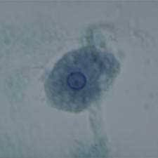
 Entamoeba histolytica cyst and trophozoite, haematoxylin stained
© Dr Peter Darben, Queensland University of Technology clinical parasitology collection. Used with permission Entamoeba histolytica trophozoites in section of intestine (H&E)
© Dr Peter Darben, Queensland University of Technology clinical parasitology collection. Used with permission
 Parasitic amoeba (Entamoeba histolytica) causes amebic dysentery & ulcers (vegetative trophozoite stage). Amebic dysentery is spread by fecal contamination of food and water and is most common where sanitation is poor.
© Dennis Kunkel Microscopy, Inc. Used with permission
CDC DPDx Parasite Image LibraryC
 Trophozoites of Entamoeba histolytica. Trichrome stain. Two diagnostic characteristics are seen here: two of the trophozoites have ingested erythrocytes, and the nuclei have typically a small, centrally located karyosome, as well as thin, uniform peripheral
chromatin.
CDC DPDx Parasite Image Library
 Entamoeba histolytica cyst and trophozoite, haematoxylin stained
© Dr Peter Darben, Queensland University of Technology clinical parasitology collection. Used with permission
 Entamoeba histolytica trophozoites in section of intestine (H&E)
© Dr Peter Darben, Queensland University of Technology clinical parasitology collection. Used with permission
 Parasitic amoeba (Entamoeba histolytica) causes amebic dysentery & ulcers (vegetative trophozoite stage). Amebic dysentery is spread by fecal contamination of food and water and is most common where sanitation is poor.
 © Dennis Kunkel Microscopy, Inc. Used with permission
Cyst
(figure 2)
A  B
B  Cysts of Entamoeba histolytica, stained with trichrome (A) and iodine (B). Each cyst has 4 nuclei, of which 3 (in A) and 2 (in B) are visible in this focal plane (the fourth nucleus is coming into focus in D). The nuclei have characteristically centrally located karyosomes. The cyst in A contains a large chromatoid body. Entamoeba histolytica cysts measure 12-15 µm
CDC DPDx Parasite Image Library
 B
B  Cysts of Entamoeba histolytica, stained with trichrome (A) and iodine (B). Each cyst has 4 nuclei, of which 3 (in A) and 2 (in B) are visible in this focal plane (the fourth nucleus is coming into focus in D). The nuclei have characteristically centrally located karyosomes. The cyst in A contains a large chromatoid body. Entamoeba histolytica cysts measure 12-15 µm
CDC DPDx Parasite Image Library
Life Cycle of Entamoeba histolytica
(figure 3)

Infection by Entamoeba histolytica occurs by ingestion of mature cysts (1) in fecally contaminated food, water, or hands. Excystation (2) occurs in the small intestine and trophozoites (3) are released, which migrate to the large intestine. The trophozoites multiply by binary fission and produce cysts (4) , which are passed in the feces. Because of the protection conferred by their walls, the cysts can survive days to weeks in the external environment and are responsible for transmission. (Trophozoites can also be passed in diarrheal stools, but are rapidly destroyed once outside the body, and if ingested would not survive exposure to the gastric environment.) In many cases, the trophozoites remain confined to the intestinal lumen (A: non-invasive infection) of individuals who are thus asymptomatic carriers and cysts passers. In some patients the trophozoites invade the intestinal mucosa (B: intestinal disease), or, through the bloodstream, extraintestinal sites such as the liver, brain, and lungs (C: extra-intestinal disease), with resultant pathologic manifestations. It has been established that the invasive and noninvasive forms represent separate species, respectively E. histolytica and E. dispar, which are morphologically indistinguishable. Transmission can also occur through fecal exposure during sexual contact (in which case not only cysts, but also trophozoites could prove infective).
Pathology
(figure 4)
 Gross pathology of liver containing amebic abscess
CDC/Dr. Mae Melvin; Dr. E. West of Mobile, AL DPDx Parasite Image Library
 Gross pathology of amebic abscess of liver. Tube of "chocolate" pus from abscess.
CDC/Dr. Mae Melvin; Dr. E. West of Mobile, AL
 Histopathology of a typical flask-shaped ulcer of intestinal amebiasis
CDC/Dr. Mae Melvin
----------------------------------------------------------------------------------------------------------------------------------------------------------------
You Tube: Watch the Video of Ameba's Motility (click on the picture):
1. 
2. 


2 komentar:
hahaha.. kacauu! smp amebiasis jg dmasukin blog :D
*curiga ini kluar pas SOOCA tan..
pas banget cha, kata2nya jadi doa tu.. jadi kasus SOCA guw, sm2 diare ;D
lumayan jadi DD-nya..hihi
Post a Comment